Case 1: Hip Fracture (Intertrochanteric Fracture)
70 Years female presented with right hip fracture following a fall in the bathroom
Images
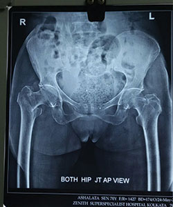
XRAY - 1
X-RAY showing right hip fracture (Intertrochanteric Fracture)/p>
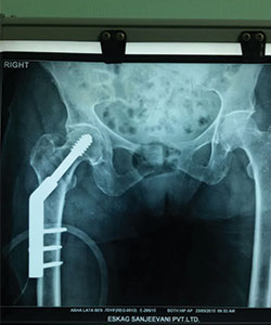
XRAY - 2
post operation X-RAY (fracture fixed with Dynamic hip screw)
Videos
Patient walking independently without support 6 weeks after surgery
Case 2: Patella Fracture
Patella fracture in a 70years old female.
Images
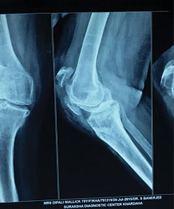
X-RAY 1
patella fracture (pre operative x-ray)
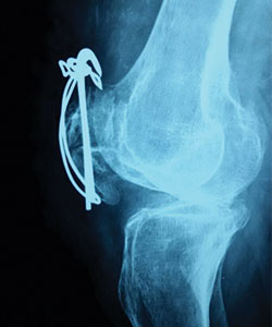
X-RAY 2
post operative x-ray of patella fracture
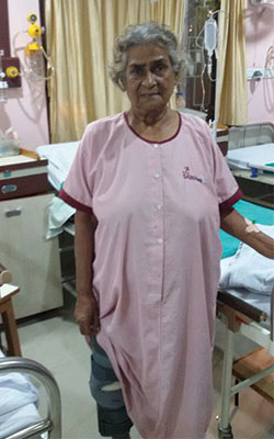
IMAGE - 1
Patient walking 24 hours after operation for patella fracture
Case 3: Comminuted hip fracture
Comminuted left hip fracture in a 81 years old male patient
Images
X-RAY 1
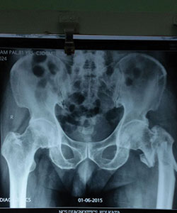
pre operative x-ray showing comminuted left hip fracture
X-RAY 2
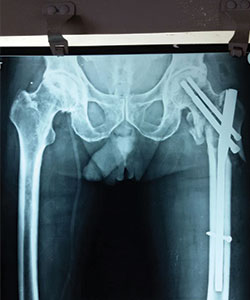
Post operative x-ray (PFN done)
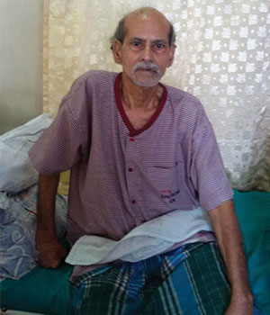
IMAGE 1
Patient is fit after operation for comminuted left hip fracture
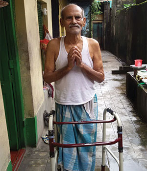
IMAGE 2
Patient is walking with walker support 4 weeks after operation
Case 4: Tibial Fracture
70 Years female fell in the bathroom and sustained injury to his left leg. She had fracture of left leg (tibia and fibula)
Images

X-RAY 1
Pre operative x-ray showing fracture both bone left leg
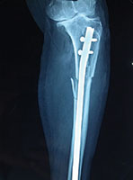
X-RAY 2
Post operative Xray
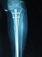
X-RAY 3
Post op xray tibial fracture fixed with interlocking nail
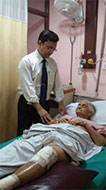
IMAGE 1
Dr Saikat with the patient
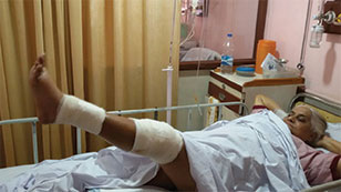
IMAGE 2
Patient actively raising operative leg 24 hours after surgery
Videos
Patient walking with support 4 weeks after surgery
Case 5: Clavicle fracture
Images
X-RAY 1
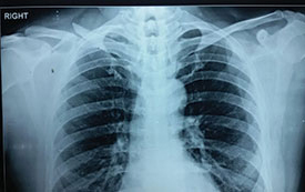
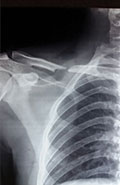
Displaced fracture right clavicle
X-RAY 1
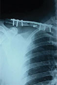
Post operative x-ray

IMAGE - 1
Patient lifting hands day after operation
Case 6: Clavicle fracture
Displayed Clavicle fracture in a 24 years old cricketer
Images
X-RAY 1
pre operative X-Ray showing displayed clavicle fracture
X-RAY 1
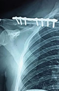
post operative x-ray showing fracture fixed with plate and screw
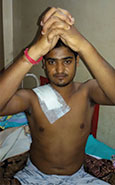
IMAGE - 1
Clavicle fracture-patient is doing exercise after operation
Case 7: Total Knee Replacement
54 years female was suffering from both knee pain for last 4-5 years. She had bilateral advanced osteoarthritis knee.
Patient underwent knee replacement surgery in two stages. 1st right knee was replaced after 8 months left knee was replaced.
Images

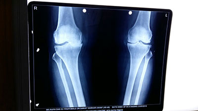
X-RAY 1
knee advanced stage of osteoarthritis

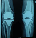
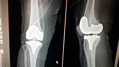
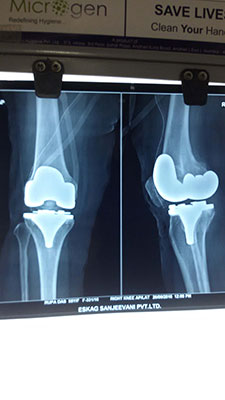
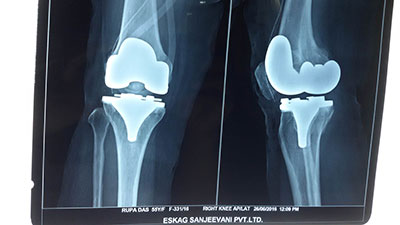
X-RAY 2
post op xray after knee replacement

X-RAY 3
xray left knee lateral view after knee replacement

IMAGE - 1
knee replacement photo
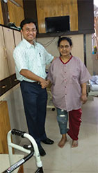
IMAGE - 2
Dr Saikat with the patient

IMAGE - 3
24 hours after left knee replacement surgery
Videos
Patient walking with severe knee pain before knee replacement
Patient walking with support 48 hours after right knee replacement
Patient walking painfree after bilateral knee replacement (1 year after surgery)
Case 8: Total Knee Replacement
Both knee osteoarthritis in a 62 years female.
Images

X-RAY 1
Both knee osteoarthritis
X-RAY 2
xray after both knee replaced
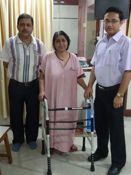
IMAGE - 1
Patient walking with support two days after surgery
Case 9: Total Knee Replacement
Images
X-RAY 1
X-RAY both knees
X-RAY 2
X-RAY both knees
X-RAY 3
X-RAY both knees
X-RAY 4
X-RAY both knees

IMAGE - 1
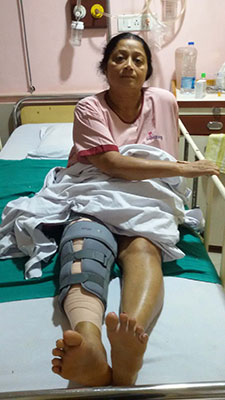
IMAGE - 2
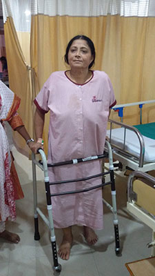
IMAGE - 3

IMAGE - 4
Videos
Patient walking with severe knee pain before operation
Patient walking painfree after both knee replacement